
Digital imaging in the service of diagnosis
Dental radiography shows what the eye cannot see: the inside of the teeth, the roots and the bone that supports them. At Cabinet Côté Gare, examinations are carried out on site, with a DEXIS panoramic and 3D (cone beam) unit installed in its own room, digital intra-oral sensors and a KaVo DIAGNOcam Vision Full HD intra-oral camera. The dentist chooses the images useful for your situation and explains them to you on screen.
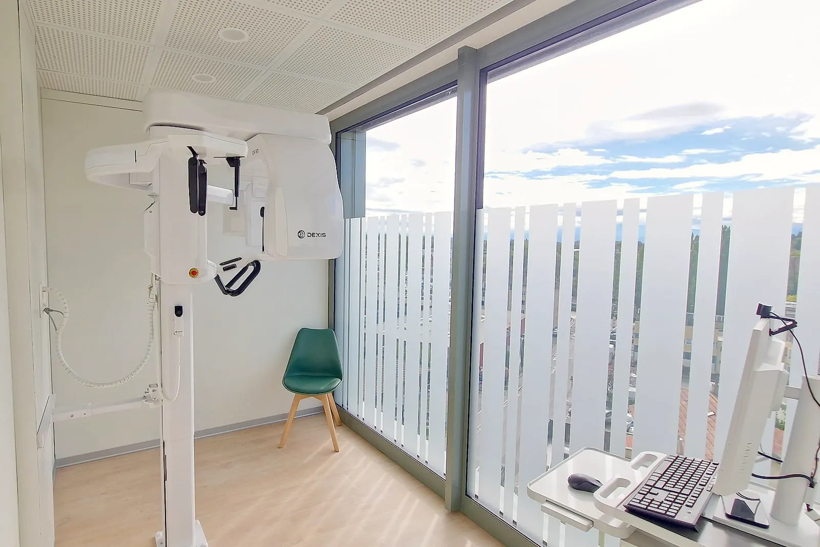
Examinations offered at the practice
- Intra-oral X-rays (periapical and bite-wing). Small images taken with a sensor placed in the mouth. They show one or two teeth in detail, the roots, or the spaces between teeth where cavities hide.
- Panoramic X-ray. A single image of both jaws: all the teeth, the wisdom teeth, the bone and the sinuses. It gives an overall view, for example for a first assessment.
- 3D imaging (CB-CT, cone beam). A three-dimensional volume of the bone and teeth, showing the course of the nerves. It is requested in complex situations, for example before placing an implant.
- KaVo DIAGNOcam Vision Full HD intra-oral camera. It works with light rather than X-rays. It complements the X-ray in detecting certain cavities between teeth and shows you the condition of a tooth on screen.
When is an X-ray useful?
The dentist requests an image when it provides information that a visual examination cannot give. The most common situations:
- detecting a cavity between two teeth or under an old restoration;
- understanding the origin of pain: infection at the tip of a root, impacted wisdom tooth, cyst;
- measuring the level of bone around the teeth during a gum assessment (periodontics) ;
- planning an implant or a bone graft : height and thickness of the bone, position of the nerves and sinuses;
- preparing a root canal treatment or a difficult extraction;
- planning an orthodontic treatment ;
- following the progress of a treatment after a few months or a few years.
What happens during the examination?
- Explanation. The dentist presents the planned images and their purpose.
- Preparation. You remove anything that could interfere with the image (earrings, necklace, glasses, removable appliance) and a protective apron is placed on you.
- Taking the images. For an intra-oral image, the sensor is placed in the mouth for a short moment. For a panoramic or 3D image, you stay still for a few seconds while the unit rotates around your head.
- Result. The image appears immediately on screen. The dentist goes through it with you and adds it to your file.
Including set-up and explanations, the examination most often takes a few minutes, depending on the images requested.
Patient safety and comfort
Digital sensors require a low dose of X-rays. Each examination must be justified: the dentist only requests an image if it could change your care, and the image is limited to the area concerned. 3D imaging is reserved for situations where it provides useful information.
Always mention a possible pregnancy before the examination. For pregnant women and for children, additional precautions are taken, and anything that can wait is postponed. The facilities comply with the radiation protection standards in force in Switzerland.
Your X-rays taken elsewhere
Bring your previous X-rays, or ask your former practice to send them to us: they make it possible to follow the progress of a tooth and avoid retaking recent images. Digital images can be easily sent to another practitioner or to a specialist when your situation requires it, with your consent.
Radiology and prices
The prices of check-up X-rays and 3D imaging are listed on the page Prices. A detailed treatment plan and a personalised quote are given to you at the first consultation, before any treatment begins.
Frequently asked questions
Are dental X-rays dangerous?
The dose from a dental X-ray is low: it is equivalent to a few hours of exposure to natural background radiation. Each examination is justified by the dentist, who limits the image to the relevant area.
How often should X-rays be taken?
According to clinical needs: full assessment every 3-5 years, targeted checks in case of pain or specific treatment.
Is cone beam always necessary?
No. It is used for complex cases: implantology, surgery, difficult endodontics or suspected pathology.
How much do X-rays cost?
Two bite-wing X-rays are CHF 49.90. A simple check-up by the dentist, at CHF 92.-, includes two check-up X-rays. 3D imaging (CB-CT) is CHF 394.-.
Can I bring X-rays taken elsewhere?
Yes, and it is useful: it avoids retaking recent images. Bring them, or ask your former practice to send them to us.
Are X-rays possible during pregnancy?
Always mention a pregnancy. A dental X-ray is very localised and a protective apron is used; anything that can wait is postponed.
What does cone beam show in addition?
A three-dimensional image of the bone volume and the course of the nerves. It is useful before an implant, a complex extraction or a difficult root canal treatment.
Are the images digital?
Yes. The digital sensor requires a lower dose than conventional film, and the image is available immediately during the consultation.
What does a panoramic X-ray show?
An overall view of both jaws on a single image: all the teeth, the wisdom teeth, the bone and the sinuses. The detail of a cavity between two teeth is checked on an intra-oral image.
What is the DIAGNOcam camera for?
It illuminates the tooth with light, without X-rays, to detect certain cavities between teeth and show you the condition of a tooth on screen. It complements the X-ray when one is indicated.
See also: The check-up with two X-rays.
